Русский
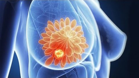
Significant changes in life conditions and lifestyle over last 100 years led to high mastopathy and breast cancer prevalence.
Mastopathy is the most common female pathology. According to different experts, the disease occurs in 30-60% of women. The term covers a range of benign dishormonal breast abnormalities manifested by diffuse induration or nodularity, breast tenderness (mastodynia), and sometimes nipple discharge. The term “dishormonal” implies underlying hormonal imbalance.
Mastopathy was described in 1838. There are many mastopathy synonyms out of nowadays use (“chronic induration”, “Schimmelbusch disease”, “chronic cystic mastitis”, “cystic serous breast tumor”, “cystic adenoma”, etc.) that have appeared due to various disease manifestations (with or without cysts, mostly local or diffuse indurations) and its consequences (various tissue alterations). Meantime, thefollowing terms are used to indicate mastopathy: fibroadenomatosis and fibrocystic breast disease.
Etiology
Mastopaty is one of the most discussed and studied diseases. The reason of is it’s role of a breast cancer biomarker. Although mastopathy does not cause breast cancer, its presence increases incidence of cancer 3-5 times as compared with general population. And proliferative mastopathy increases breast cancer risk 25-30 times.
One of the main causes of so high mastopathy and breast cancer prevalence rate is changed “gynecological portrait” of a woman in developed countries:
- early menarche (12-14 years),
- menopause at the age of 50-55,
- giving birth to 1-2 children,
- short period of breast feeding (4-6 months),
- increased reproductive age to 40 years,
- quadripled number of menstrual cycles (up to 400),
- significant increase in a number of abortions.
Mastopathy symptoms
Complains of pain in the breast, various in character (sharp, burning, stabbing) and intensity (mild pain in pain hypersensitivity, severe pain up to megalgia). Pain feeling and discomfort grow stronger before menstruation.
Small nodules or breast indurations with hardly palpable borders that can be detected by the woman or by her mammologist.
axillary lymph-node hyperplasia, sometimes hypersensitivity inpressing.
Breast enlargement
Nipple discharge (strong or poor, in palpation)
You can examine your breast on your own (video)
There are several mastopathy classifications accepted by the global community. One of the most competent and full classifications is a clinical one, according to which there are 3 forms of mastopathy distinguished:
Mastalgia (mastodynia) – manifests by painful feelings in the breast caused by pressing (complain of pain in the breast), sometimes by beast deformation such as change of size, form and structure (induration, swelling).
Diffuse mastopathy – its main symptom is formation of breast indurations and cysts. There are 2 types of diffuse mastopathy:
- Diffuse fibrous mastopathy is characterized by prevalence of sclerosis (indurations appear in connective breast tissues);
- Diffuse fibrocystic mastopathy takes place when cysts are diagnosed with underlying sclerosis.
Localized mastopathy (localized fibroadenomatosis)
Localized mastopathy is characterized by flexible flipperlike indurations that are painful in touch. If the induration has distinct borders and can be called dense, it is a cyst.
To rule out breast cancer, a mammologist examines mammary glands and recommends mammography followed by assessment investigation of detected nodule cells and tissues.
It happens that tumor etiology can be only identified surgically. In this regard it is necessary to conduct partial mastectomy of suspicious breast tissue. The removed breast tissue is being assessed histologically (tissues are frozen, the surgery team waits for comments from the laboratory). The result may indicate localized fibroadenomatosis or breast cancer (in this case surgery goes on, the mammary gland is removed).
Mammary gland fibroadenoma is a form of localized mastopathy that generally occurs to youths (it is worth noting that pediatric mastopathy doesn’t exist, but it can occur in girls of adolescent and puberty age). It is diagnosed by the mammalogcal examination in case palpable movable indurations are detected. These indurations are usually smooth and round in touch. There is also the other term of fibroadenomas in medical litearature – “breast mouse”.
One more classification was proposed in 1985, it distinguishes 2 forms of mastopathy according to the clinical and radiographic signs:
Diffuse mastopathy:
a) with glandular dominant (adenosis)
b) with fibrous dominant (fibrosis)
c) with cystic dominant (multiple cysts)
d) mixed form (cystic glandular)
Nodal mastopathy:
a) fibroadenoma
b) cyst
It is worth noting that other disease term can be met in modern medical practice, which are full or partial synonyms of mastopathy:
- Fibroadenomatosis (do not confound with fibroadenoma)
- Cystic mastopathy (full synonym of fibrocystic breast – FCB)
- Cystic fibroadenomatosis (the same as FCB)
- Cystic disease (used in case of cystic dominant)
- Mints disease (used in case of breast ducts papillomatosis)
- Adenosis
- Dishormonal hyperplasia
- Mammary dysplasia
- Fibrocystic mastopathy
FIBROCYSTIC MASTOPATHY – THERAPY AND PREVENTION
Fibrocystic mastopathy (fibrous mastopathy) is a group of breast diseases characterized by altered morphology of glandular and connective tissue provoking alternation of breast blood supply, congestion and cyst fromation. Average “age of disease” is 30 to 50 years.
Etiology of fibrocystic mastopathy
- Ovarian dishormonal disorders (with elevated level of estrogen and decreased level of progesterone);
- Ovary inflammation;
- Thyroid or other dyssecretion;
- Hepatic diseases (hepatitis, cirrhosis) with disorder of steroid metabolism;
Symptoms of cystic mastopathy
- Breast pain (dull, aching pain appeared when pressing and squeezing breasts) intensified before menstruation.
- Indurations palpable in careful breast examination. They appear in the middle and the second half of the menstrual cycle, and decrease or disappear completely as the menstruation begins. Moreover, breasts swell, their weight and volume increase without obvious reasons. Sometimes lymphatic nodes swell. Woman can examine her breasts on her own, ideally on menstrual cycle day 7-10. The Woman should place one hand behind her head and palpate the breast on the other side with the other hand. Mostly often, indurations locate in the upper-external quadrant of any breast.
- If the disease lasts for long, the woman can notice nipple discharge. Its color may vary from whitish to green and brownish. Any blood seen is direct indication for urgent consultation with the oncologist-mammologist.
Only a mammologist may prescribe therapy for fibrocystic mastopathy after complete examination of the woman. AS a rule, the mammologist refers hormone tests, radiological assessment (mammography), and US breast scan.
Quite often fibrocystic mastopathy disappears spontaneously during menopause. In more complicated cases, when doctor’s attention is required, the main target of treatment is normal hormonal levels and pain minimizing or alleviation. As a rule, beside analgesics, the doctor prescribes vitamins and special medicines to treat mastopathy.
Up to date, only four special medications are registered for therapy of mastopathy in the Russian Federation. One of them is MAMOCLAM.