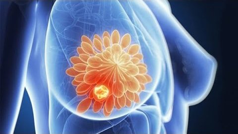
Mammary gland investigation using the ultrasound equipment is called breast echography.
It is generally carried out using high resolution linear probes (high resolution provided by the high emission frequency – 7-10 MHz) of the modern equipment.
The conducted examination allows for:
- detecting and measuring the volume of breast cysts: the cyst looks like an oval or round formation with even and thin capsule and clear margins. Ultrasound investigation allows detecting the cyst even if its size does not exceed 2-3 mm. Besides, ultrasonography specialist can define the cyst type, for it can be simple (benign) or atypical (possibly malignant, which is defined by the presence of tissue mass and hemorrhagic content, indicating the necessity of further tests to diagnose cancer).
- assessing of the axillary and other adjacent lymphatic nodes in order to reveal signs of malignant tumor spreading in them.
- detecting the tumor of the mammary glands (fibroadenoma, cancer node).
- detecting the dilated lactiferous ducts.
The fixation of revealed abnormalities is carried out using thermal paper and a special printer. The echography results are needed to monitor the dynamics of Mastopathy and counted during repeated visits to mammologist.
Of note – echography is harmless (it does not include exposure any radiation of the mammary glands), and it provides lots of useful information, which is why specialist recommend periodical self-examinations.


 Русский

